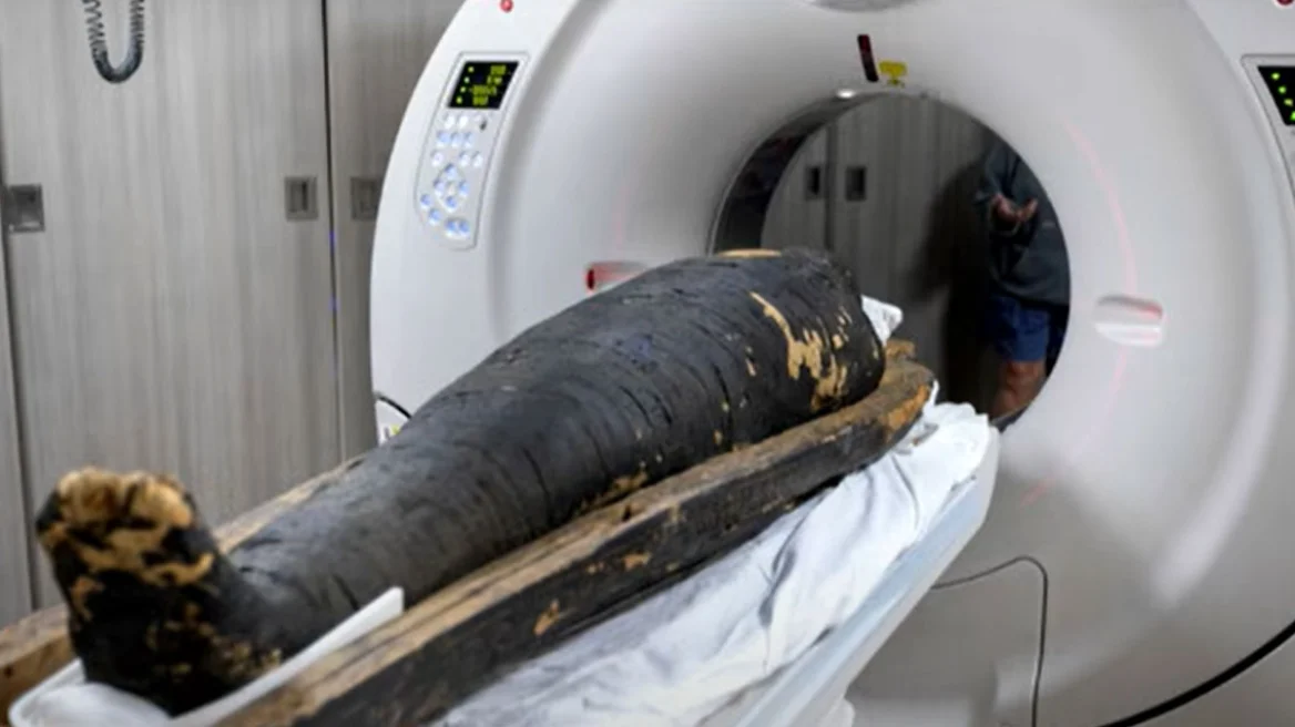
Radiologists from Keck Medicine of USC used high-resolution computed tomography (CT) scans to examine two ancient Egyptian mummies, offering an unprecedented look at human bodies preserved for more than two thousand years. The research focused on two priests: Nes Min, dated to around 330 BC, and Nes Hor, from approximately 190 BC. Each body was scanned while still resting in the lower section of a heavy sarcophagus weighing about 90 kilograms.
The CT scanner captured magnificent slices throughout the entire body. Specialists then combined hundreds of individual images into three-dimensional digital models. These models revealed details rarely visible in mummies, such as eyelids, lips, and the structure of the facial bones, allowing a more immediate sense of the individual appearance of the people behind the wrappings.
The scans also recorded signs of aging and disease. Nes Min showed severe damage in the lower spine, with collapse of a lumbar vertebra—an image associated with long-term strain and degenerative wear, similar to what is seen today in elderly patients with chronic lower back pain. Nes Hor displayed a different set of problems: advanced tooth decay in several teeth and significant wear in one hip joint, damage that would likely have caused pain and difficulty walking. The overall condition of the bones indicates that Nes Hor reached an older age at death compared to Nes Min.
The images also captured burial objects. Nes Min had been buried with small items shaped like a scarab and a fish, placed within the wrappings and unseen for more than 2,000 years. The CT data allowed these objects to be measured and studied without any physical intervention on the mummy.
After imaging, visualization specialists created digital reconstructions of the skeletons and selected findings. Full-scale physical prints of skulls, spinal columns, hips, and burial objects were then produced using medical-grade 3D printers. These replicas will be presented to the public at the California Science Center, alongside the mummies themselves and digital applications, as part of the exhibition “Mummies of the World,” which opens on February 7.
The same imaging and 3D-printing process is widely used in modern medicine. Hospitals use CT or MRI scans to create digital 3D models of organs such as the liver, heart, or pelvis, which surgeons study before complex operations. Physical replicas help plan incisions, choose implants, and practice critical steps. A key role in this work is played by the USC Center for Innovation in Medical Visualization, which operates nearly two dozen printers serving both clinical practice and research projects, including the study of the mummies.
According to doctors, allowing patients to interact with a physical 3D model helps them better understand their anatomy and the proposed procedures, improving communication. This research program demonstrates how technologies designed to care for living patients can also illuminate the lives of people from antiquity—preserving fragile remains intact while revealing insights into health, injuries, and daily life in ancient Egypt.
Ask me anything
Explore related questions